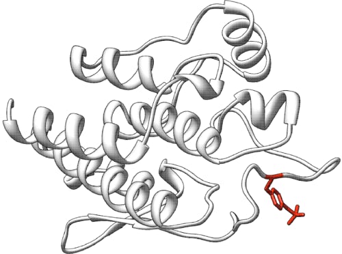
Pericentriolar Material 1 (PCM1): predict the effects of missense changes in a zebrafish model
Challenge: PCM1 missense
Variant data: public
Last updated: 14 April 2018
This challenge is closed.
Make sure you understand our Data Use Agreement and Anonymity Policy
Summary
The PCM1 (Pericentriolar Material 1) gene is a component of centriolar satellites occurring around centrosomes in vertebrate cells. Several studies have implicated PCM1 variants as a risk factor for schizophrenia. Ventricular enlargement is one of the most consistent abnormal structural brain findings in schizophrenia Therefore 38 transgenic human PCM1 missense mutations implicated in schizophrenia were assayed in a zebrafish model to determine their impact on the posterior ventricle area. The challenge is to predict whether variants implicated in schizophrenia impact zebrafish ventricular area.
Background
The PCM1 (Pericentriolar Material 1) gene is a component of centriolar satellites occurring around centrosomes in vertebrate cells. PCM1 can form a complex at the centrosome with DISC1 protein. In addition to containing centrosomal proteins, the centriolar satellites can move along microtubules. The centrosomal proteins help anchor the microtubules at the centrosome. The centrosome is essential for neurodevelopment (specifically cortical development). If PCM1 is mutated, localization of centrosomal proteins may be altered (not correctly localized). Thus, certain components of the PCM1 have been implicated in genetic susceptibility to cancers, schizophrenia, and other mental diseases (Kamiya et al., 2008; Zoubovsky et al., 2015).
Several studies have implicated PCM1 variants as a risk factor for schizophrenia. PCM1 mutant mice have shown behavioral deficits (Moens et al., 2010). However, other studies have found low risk or no association (Zoubovsky et al., 2015).
Experiment
The Katsanis lab assessed 38 missense mutations within PCM1 in a zebrafish model.
Native zebrafish embryo PCM1 protein was suppressed by injecting morpholino (MO) antisense oligonucleotides to inhibit translation of mRNA of the pcm1 gene. MOs are stable molecules that consist of a large, nonribose morpholine backbone with the four DNA bases pairing stably with mRNA at either the translation start site (to disrupt protein synthesis) or at intron-exon boundaries (to disrupt mRNA splicing) (Summerton & Weller, 1997). Morpholinos have been shown to bind to and block translation of mRNA in vitro, in tissue culture cells, and, in vivo (Davis et al., 2014). Embryos deficient in pcm1 function show an absence of brain ventricle formation.
For each mutation, the Katsanis lab injected a group of embryos with MO and the mRNA of the human gene carrying the mutation (MO+VAR). The brain ventricle formation of the group of the (MO+VAR) animals was compared to the brain ventricle formation measured in a group of animals with MO alone and a group with MO+WT. In order to access the effect on mutations on ventricle size, the ventricle space is filled with a fluorescent dye and imaged by brightfield and fluorescent microscopy (Niederriter et al., 2013; Gutzman & Sive, 2009). Images were taken (Figure 1). To estimate the ventricle structure, an automated image processing tool was applied to quantitate its volume (Mikut et al., 2013; Naslund, 2014). P values for statistically different volume of brain ventricle between pairs of conditions (Lowery et al., 2009) were obtained using a Student’s t-test. When the p-value is:
- NOT statistically different from MO, but statistically significantly different from MO+WT, the variant is pathogenic or loss of function.
- Statistically different from MO, but not from MO+WT, the variant is benign
- Statistically different from MO, and at the same time statistically significantly different from MO+W, the variant is hypomorphic or partially loss of function.

Figure 1: Example of brain ventricle comparison (taken from Lowery et al., 2009). Ventricles in gures B and C show an abnormal midbrain-hindbrain boundary compared to WT (A). On the other hand, ventricles E and F show regions mostly recovered compared to WT (D).
Prediction challenge
Participants are asked to submit the probability of the effect of the variants on zebrafish brain development.
Download dataset: 5- PCM1_dataset.txt
Download submission template: 5-PCM1_template_v02.txt
Download validation script: 5-PCM1_validation_script.pl and 5-PCM1_cagi-validate_v02.R
Prediction submission format
The prediction submission is a tab-delimited text file. Organizers provide a file template, which must be used for submission. In addition, a validation script will be provided, and predictors must check the correctness of the format before submitting their predictions. In the submitted file, each row should include the following tab-separated fields:
- Nucleotide position: DNA coding change of the variant (e.g., c.G17A) on the NM_001315507 transcript.
- Variant: protein change of the variant (e.g. p.G6D), on UniProtKB protein Q15154 (PCM1_HUMAN)
- p-value relative of change from MO: The probability that this variant is statistically different from MO.
- Standard deviation: This defines the confidence of the prediction in column 3. Large SD means low confidence, while small SD means that the predictor is confident about the submitted prediction.
- p-value of change from MO+WT: The probability that this variant is statistically different from MO+WT
- Standard deviation: This defines the confidence of the prediction in column 5. Large SD means low confidence, while small SD means that the predictor is confident about the submitted prediction.
- Functional effect: pathogenic (2), hypomorphic (1), or benign (0)
- Confidence: in the functional effect assignment ranges 0.0 to 1.0. (1.0 implies total confidence in the assignment)
- Comments: Optional brief comments based on the predictions.
In the template file, cells in columns 3-9 are marked with a "*". Submit your predictions by replacing the "*" with your value. No empty cells are allowed in the submission. If you choose not to enter a prediction leave the "*" in those cells. If you are not confident in a prediction for an individual, enter a large standard deviation for the prediction. Optionally, enter brief comments indicating the basis of the predictions; otherwise, leave the "*" in these cells. Please make sure you follow the submission guidelines strictly.
In addition, your submission must include a detailed description of the method used to make the predictions, similar in style to the Methods section in a scientific article. This information will be submitted as a separate file.
To submit predictions, you need to create or be part of a CAGI User group. Submit your predictions by accessing the link "All submission forms" from the front page of your group. For more details, please read the FAQ page.
Ethical approval
All animal experiments were approved by the Duke University Institutional Care and Use Committee.
Dataset provided by
Maria Kousi, Nicholas Katsanis, Duke University
References
Davis EE, et al. Interpreting human genetic variation with in vivo zebrafish assays. Biochim Biophys Acta (2014) 1842(10):1960-1970. PubMed
Gutzman JH, Sive H. Zebrafish brain ventricle injection. J Vis Exp (2009) 26:1218. PubMed
Kamiya A, et al. Recruitment of PCM1 to the centrosome by the cooperative action of DISC1 and BBS4: a candidate for psychiatric illnesses. Arch Gen Psychiatry (2008) 65(9):996-1006. PubMed
Lowery LA, et al. Characterization and classification of zebrafish brain morphology mutants. Anat Rec (2009) 292(1):94-106. PubMed
Moens LN, et al. PCM1 and schizophrenia: a replication study in the Northern Swedish population. Am J Med Genet B Neuropsychiatr Genet (2010) 153B(6):1240-1243. PubMed
Niederriter AR, et al. In vivo modeling of the morbid human genome using Danio rerio. J Vis Exp (2013) 78:e50338. PubMed
Summerton J, Weller D. Morpholino antisense oligomers: design, preparation, and properties. Antisense Nucleic Acid Drug Dev (1997) 7(3):187-195. PubMed
Zoubovsky S, et al. Neuroanatomical and behavioral deficits in mice haploinsufficient for pericentriolar material 1 (pcm1). Neurosci Res (2015) 98:45-49. PubMed
Revision history
9 November 2017: Initial release
9 November 2017: Details regarding the experiment and more details on submission format added
29 March 2018: New submission template
18 April 2018: New validation script
19 April 2018: Challenge closed
24 September 2018: Dataset availability added
